

A lump or abnormal growth can naturally cause concern, but not every tumor is cancerous. A benign tumor is a growth that does not invade nearby tissues or spread to distant parts of the body. However, determining whether a growth is benign requires proper medical evaluation rather than relying on symptoms or appearance alone.
Benign Tumor Diagnosis in Srinagar may involve a physical examination, imaging such as ultrasound, CT or MRI, laboratory tests, and, when appropriate, a biopsy. Imaging can identify the location and characteristics of a mass, but it cannot always establish whether a growth is benign or malignant. A tissue or cell sample may therefore be needed for a definitive diagnosis.
For patients requiring image-guided evaluation or minimally invasive treatment, Dr Irshad Banday, Interventional Radiologist (Vascular) in Srinagar, provides procedures including FNAC biopsy, embolisation, laser ablation and radiofrequency ablation (RFA), depending on the condition and clinical assessment.
Understanding Benign Tumors, Symptoms and Causes
What is a benign tumor?
A benign tumor is a non-cancerous growth. Unlike malignant tumors, benign tumors generally do not invade surrounding tissues or spread to other parts of the body. They can develop in different organs and tissues, and their symptoms depend largely on their size, location and relationship with nearby structures.
Some benign growths remain small and cause no symptoms, while others may become noticeable because of their size or location. A growth close to nerves, blood vessels or other organs can sometimes produce symptoms even when it is non-cancerous.
Common symptoms
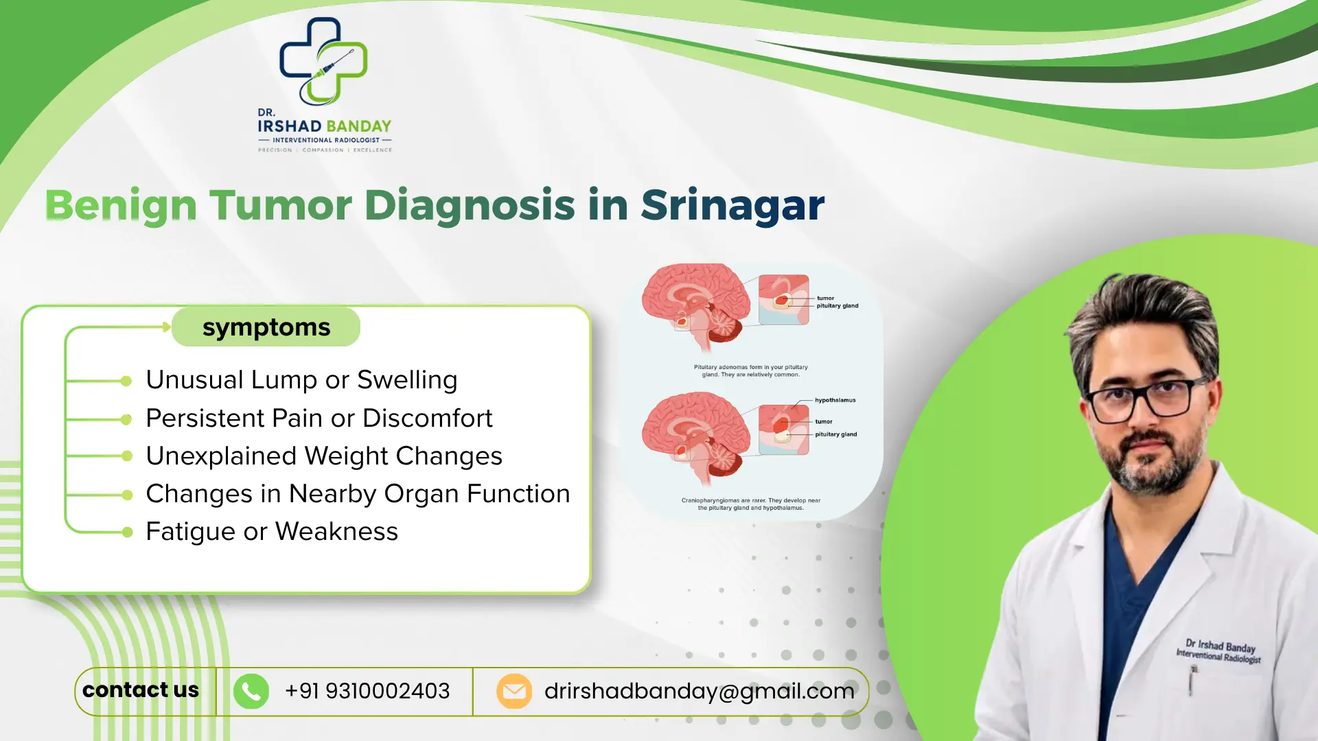
Symptoms vary according to the location and type of growth. Possible signs include:
- A new lump or swelling
- A lump that gradually increases in size
- Localised pain or discomfort
- Pressure or fullness in a particular area
- Changes in swallowing or breathing when a growth is located near the neck
- Unexplained changes in organ function
- Symptoms caused by compression of nearby structures
Importantly, these symptoms do not automatically indicate a benign tumor or cancer. Persistent or unexplained symptoms should be evaluated by a qualified doctor.
What causes benign tumors?
The exact cause varies considerably between tumor types. Some benign growths are associated with genetic or cellular changes, while others may develop without an identifiable cause. Certain conditions can also increase the likelihood of developing particular types of benign growth.
Because the cause cannot always be determined from symptoms alone, medical assessment is important when a new or changing lump is detected.
Benign Tumor Diagnosis in Srinagar can help determine the nature of an abnormal growth and guide the next step in management.
How Is a Benign Tumor Diagnosed?
Medical examination and history
The diagnostic process usually begins with a discussion of symptoms, medical history and any noticeable changes in the growth. The doctor may examine the affected area to assess its size, consistency, mobility and tenderness.
The exact diagnostic approach depends on the suspected location and type of lesion.
Imaging tests
Imaging can provide important information about the size, location and characteristics of a growth. Depending on the clinical situation, doctors may recommend:
- Ultrasound: Often useful for evaluating soft-tissue lumps and nodules.
- CT scan: Provides detailed cross-sectional images and can help assess deeper structures.
- MRI: Offers detailed soft-tissue imaging and can help define the relationship between a lesion and surrounding structures.
- Other specialised imaging: May be considered depending on the suspected condition.
Imaging can help doctors decide whether surveillance, further testing or tissue sampling is appropriate. However, imaging alone cannot always distinguish a benign lesion from a malignant one.
FNAC and biopsy
A biopsy involves collecting cells or tissue from an abnormal area for laboratory examination. Fine-needle aspiration cytology (FNAC) uses a thin needle to obtain cells for analysis. Image guidance can help the doctor accurately target the area being sampled.
For example, ultrasound-guided thyroid FNAC uses real-time ultrasound to guide a fine needle into a thyroid nodule. The collected cells are then examined by a pathologist.
Image-guided needle biopsy is generally less invasive than an open surgical biopsy and is commonly performed as an outpatient procedure.
Understanding the pathology report
After FNAC or biopsy, a pathologist examines the sample and prepares a report. The result may help determine whether the lesion is benign, malignant, suspicious or requires additional evaluation.
The final treatment decision should consider the pathology result together with imaging findings, symptoms and the patient’s overall clinical situation.
Benign Tumor Diagnosis in Srinagar therefore involves more than identifying a lump; it is about establishing an accurate diagnosis before deciding whether treatment or observation is appropriate.
Benign Tumor Treatment, Minimally Invasive Procedures and Recovery
When does a benign tumor need treatment?
Not every benign tumor requires immediate treatment. If a lesion is small, asymptomatic and considered low-risk, a doctor may recommend observation with periodic clinical assessment or imaging.
Treatment may be considered when a benign growth:
- Causes pain or discomfort
- Produces pressure symptoms
- Affects normal organ function
- Continues to grow
- Causes cosmetic or functional concerns
- Has imaging or pathological features requiring further management
The appropriate approach depends on the specific diagnosis.
Benign Tumor Treatment options
Treatment can include observation, medication in selected conditions, surgery, or minimally invasive procedures. The choice should be based on the tumor type, location, size, symptoms and available treatment options.
For selected lesions, interventional radiology may offer image-guided treatment without the need for a large surgical incision.
Dr Irshad Banday provides interventional procedures in Srinagar that may include:
- FNAC Biopsy: Image-guided sampling of cells from selected nodules or lesions for diagnostic evaluation.
- Embolisation: A minimally invasive procedure that intentionally blocks selected blood vessels supplying a target area when clinically appropriate.
- Laser Ablation: Uses laser energy to heat and destroy selected abnormal tissue under image guidance.
- Radiofrequency Ablation (RFA): Uses radiofrequency energy to generate controlled heat within a targeted lesion.
- Vascular Interventions: Image-guided procedures for selected vascular conditions.
These procedures are not appropriate for every benign tumor. A detailed evaluation is necessary to determine whether an intervention is suitable.
Benefits of image-guided minimally invasive treatment
When appropriate for a particular condition, minimally invasive image-guided procedures may offer several potential advantages compared with conventional open surgery, such as:
- Smaller access point rather than a large incision
- Image guidance for accurate targeting
- Potentially shorter recovery for selected procedures
- Reduced disruption to surrounding tissues
- Treatment tailored to the location and characteristics of the lesion
The actual benefits and risks vary according to the procedure and individual patient.
Recovery after treatment
Recovery depends on the procedure performed, the location of the lesion and the patient’s general health.
After a needle biopsy, patients may experience temporary tenderness or bruising around the treatment site. Ultrasound-guided thyroid FNAC, for example, generally has a short recovery period, with patients often able to return to normal activities soon afterward.
After ablation or embolisation, the doctor provides specific instructions regarding activity, medication, wound care and follow-up. Follow-up imaging may be recommended to evaluate treatment response or monitor the lesion over time.
Benign Tumor Diagnosis in Srinagar should always be followed by an individualized management plan based on the confirmed diagnosis.
Diagnosis versus treatment: what is the difference?
| Diagnostic approach | Purpose |
| Physical examination | Evaluates the lump and symptoms |
| Ultrasound | Assesses structure and characteristics |
| CT/MRI | Provides detailed anatomical information |
| FNAC | Obtains cells for laboratory examination |
| Biopsy | Obtains tissue for pathological evaluation |
| Follow-up imaging | Monitors stability or changes |
A diagnostic procedure determines what the lesion may be, while a treatment procedure aims to manage the confirmed condition.
Frequently Asked Questions About Benign Tumor Diagnosis
1. Is every tumor cancerous?
No. A benign tumor is non-cancerous and generally does not invade nearby tissues or spread to distant parts of the body.
2. How is a benign tumor diagnosed?
Diagnosis may involve physical examination, imaging tests and, when necessary, FNAC or biopsy. The appropriate combination depends on the location and characteristics of the lesion.
3. Can ultrasound identify a benign tumor?
Ultrasound can provide useful information about many lumps and nodules and may show features that suggest a benign lesion. However, it cannot always establish the diagnosis with certainty, so additional testing may sometimes be required.
4. What is FNAC?
FNAC, or fine-needle aspiration cytology, uses a thin needle to collect cells from a lump or nodule. The cells are examined in a laboratory to help establish a diagnosis.
5. Is FNAC a surgical procedure?
FNAC is a minimally invasive needle procedure rather than open surgery. Image guidance, such as ultrasound, may be used to improve targeting of the lesion.
6. Does every benign tumor require removal?
No. Some benign tumors can be monitored if they are small, stable and not causing problems. Treatment may be considered if the growth causes symptoms, enlarges or requires intervention for other clinical reasons.
7. What is the role of an interventional radiologist?
An interventional radiologist uses medical imaging to guide minimally invasive diagnostic and therapeutic procedures. Depending on the condition, this can include image-guided biopsy, embolisation and ablation procedures.
8. Can benign tumors be treated without open surgery?
Some selected benign lesions may be treated using minimally invasive techniques such as ablation or embolisation. Suitability depends on the specific lesion and the patient’s clinical assessment.
9. When should I see a doctor about a lump?
A new, growing, painful or persistent lump should be medically evaluated. Early assessment can help determine whether observation, further testing or treatment is appropriate.
Conclusion: Expert Evaluation for Benign Tumors in Srinagar
A benign tumor does not mean that a growth should automatically be ignored. Correct diagnosis is important because management depends on the tumor’s location, size, symptoms, imaging characteristics and pathology results. In selected cases, minimally invasive procedures can provide diagnostic or therapeutic options while avoiding some of the tissue disruption associated with open surgery.
Benign Tumor Diagnosis in Srinagar can involve advanced imaging, FNAC or biopsy followed by an individualized treatment plan. Dr Irshad Banday, an Interventional Radiologist (Vascular) in Srinagar, provides image-guided procedures including FNAC biopsy, embolisation, laser ablation and radiofrequency ablation (RFA) for appropriately selected patients.
If you have a newly detected lump, nodule or suspected benign lesion, consult a qualified specialist for proper evaluation rather than relying on symptoms alone.
Book an appointment with Dr Irshad Banday in Srinagar for expert evaluation and image-guided diagnostic or minimally invasive treatment options.